at the CNIO

Category: Publications (Page 11 of 19)

Resident macrophage-dependent immune cell scaffolds drive anti-bacterial defense in the peritoneal cavity

Adrián Vega-Pérez, Laura H. Villarrubia, Cristina Godio, Alejandra Gutiérrez-González, Lidia Feo-Lucas, Margarita Ferriz, Natalia Martínez-Puente, Julieta Alcaín, Alfonso Mora, Guadalupe Sabio, María López-Bravo, Carlos Ardavín.

Peritoneal immune cells reside unanchored within the peritoneal fluid in homeostasis. Here, we examined the mechanisms that control bacterial infection in the peritoneum using a mouse model of abdominal sepsis following intraperitoneal Escherichia coli infection.
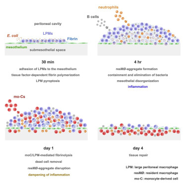
Whole-mount immunofluorescence and confocal microscopy of the peritoneal wall and omentum revealed that large peritoneal macrophages (LPMs) rapidly cleared bacteria and adhered to the mesothelium, forming multilayered cellular aggregates composed by sequentially recruited LPMs, B1 cells, neutrophils, and monocyte-derived cells (moCs). The formation of resident macrophage aggregates (resM?-aggregates) required LPMs and thrombin-dependent fibrin polymerization. E. coli infection triggered LPM pyroptosis and release of inflammatory mediators. Resolution of these potentially inflammatory aggregates required LPM-mediated recruitment of moCs, which were essential for fibrinolysis-mediated resM?-aggregate disaggregation and the prevention of peritoneal overt inflammation.

Thus, resM?-aggregates provide a physical scaffold that enables the efficient control of peritoneal infection, with implications for antimicrobial immunity in other body cavities, such as the pleural cavity or brain ventricles

Protocol for the assessment of mTOR activity in mouse primary hepatocytes

Ana Belén Plata-Gómez, María Crespo, Celia de la Calle Arregui, Lucía de Prado-Rivas, Guadalupe Sabio, Alejo Efeyan.

We present a protocol for measuring the activity of the mechanistic target of rapamycin (mTOR) pathway in ex vivo isolated mouse primary hepatocytes. It can be used as a tool for genetic, pharmacological, metabolomic, and signal transduction procedures.

We discuss critical aspects for improving yield, viability, and modulation of the mTOR pathway. This protocol can be adapted to other signaling cascades and is compatible with multiple readouts.

Inhibition of ATG3 ameliorates liver steatosis by increasing mitochondrial function

Natáliada Silva Lima, Marcos F. Fondevila, Eva Nóvoa, Xabier Buqué, Maria Mercado-Gómez, Sarah Gallet, Maria J. González-Rellan, Uxia Fernandez, Anne Loyens, Maria Garcia-Vence, Maria del Pilar Chantada-Vazquez, Susana B. Bravo, Patricia Marañon, Ana Senra, Adriana Escudero, Magdalena Leiva, Diana Guallar, Miguel Fidalgo, Pedro Gomes, Marc Claret, Guadalupe Sabio, Marta Varela-Rey, Teresa C. Delgado, Rocio Montero-Vallejo, Javier Ampuero, Miguel López, Carlos Diéguez, Laura Herrero, Dolors Serra, Markus Schwaninger, Vincent Prevo, Rocio Gallego-Duran, Manuel Romero-Gomez, Paula Iruzubieta, Javier Crespo, Maria L. Martinez-Chantar, Carmelo Garcia-Monzon, Agueda Gonzalez-Rodriguez, Patricia Aspichueta & Ruben Nogueiras.

BACKGROUND & AIMS: Autophagy-related gene 3 (ATG3) is an enzyme mainly known for its actions in the LC3 lipidation process, which is essential for autophagy. Whether ATG3 plays a role in lipid metabolism or contributes to nonalcoholic fatty liver disease (NAFLD) remains unknown.

METHODS: By performing a liver proteomic analysis from mice with genetic manipulation of hepatic p63, a regulator of fatty acid metabolism, we identified ATG3 as a new target downstream of p63. ATG3 was evaluated in liver samples of patients with NAFLD. Further, genetic manipulation of ATG3 was performed in human hepatocyte cell lines, primary hepatocytes and in the liver of mice.

JNK1 inhibitor SP600125 blunted increased lipid content (Image: Magdalena Leiva).

RESULTS: ATG3 expression is induced in the liver of animal models and patients with NAFLD (both steatosis and NASH) compared with those without liver disease. Moreover, genetic knockdown of ATG3 in mice and human hepatocytes ameliorates p63- and diet-induced steatosis, while its overexpression increases the lipid load in hepatocytes. The inhibition of hepatic ATG3 improves fatty acid metabolism by reducing c-Jun N-terminal protein kinase 1 (JNK1), which increases sirtuin 1 (SIRT1), carnitine palmitoiltransferase I (CPT1a), and mitochondrial function. Hepatic knockdown of SIRT1 and CPT1a blunts the effects of ATG3 on mitochondrial activity. Unexpectedly, these effects are independent of an autophagic action.

CONCLUSIONS: Collectively, these findings indicate that ATG3 is a novel protein implicated in the development of steatosis.

« Older posts Newer posts »

© 2026 Sabio lab

Theme by Anders Norén — Up ↑