at the CNIO

Tag: macrophages

Resident macrophage-dependent immune cell scaffolds drive anti-bacterial defense in the peritoneal cavity

Adrián Vega-Pérez, Laura H. Villarrubia, Cristina Godio, Alejandra Gutiérrez-González, Lidia Feo-Lucas, Margarita Ferriz, Natalia Martínez-Puente, Julieta Alcaín, Alfonso Mora, Guadalupe Sabio, María López-Bravo, Carlos Ardavín.

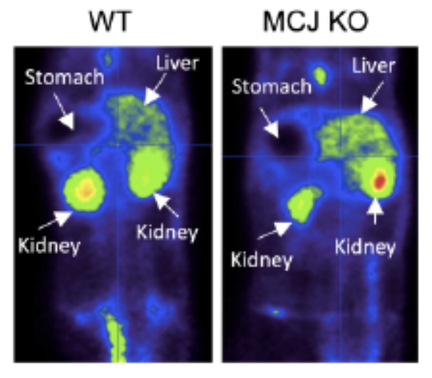
Peritoneal immune cells reside unanchored within the peritoneal fluid in homeostasis. Here, we examined the mechanisms that control bacterial infection in the peritoneum using a mouse model of abdominal sepsis following intraperitoneal Escherichia coli infection.

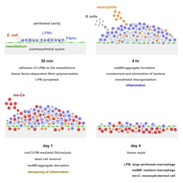
Whole-mount immunofluorescence and confocal microscopy of the peritoneal wall and omentum revealed that large peritoneal macrophages (LPMs) rapidly cleared bacteria and adhered to the mesothelium, forming multilayered cellular aggregates composed by sequentially recruited LPMs, B1 cells, neutrophils, and monocyte-derived cells (moCs). The formation of resident macrophage aggregates (resMφ-aggregates) required LPMs and thrombin-dependent fibrin polymerization. E. coli infection triggered LPM pyroptosis and release of inflammatory mediators. Resolution of these potentially inflammatory aggregates required LPM-mediated recruitment of moCs, which were essential for fibrinolysis-mediated resMφ-aggregate disaggregation and the prevention of peritoneal overt inflammation.

Thus, resMφ-aggregates provide a physical scaffold that enables the efficient control of peritoneal infection, with implications for antimicrobial immunity in other body cavities, such as the pleural cavity or brain ventricles

Mitochondrial bioenergetics boost macrophages activation promoting liver regeneration in metabolically compromised animals

Naroa Goikoetxea-Usandizaga, Marina Serrano-Maciá, Teresa C. Delgado, Jorge Simón, David Fernández Ramos, Diego Barriales, Maria E. Cornide, Mónica Jiménez, Marina Pérez-Redondo, Sofia Lachiondo-Ortega, Rubén Rodríguez-Agudo, Maider Bizkarguenaga, Juan Diego Zalamea, Samuel T. Pasco, Daniel Caballero-Díaz, Benedetta Alfano, Miren Bravo, Irene González-Recio, Maria Mercado-Gómez, Clàudia Gil-Pitarch, Jon Mabe, Jordi Gracia-Sancho, Leticia Abecia, Óscar Lorenzo, Paloma Martín-Sanz, Nicola G. A. Abrescia, Guadalupe Sabio, Mercedes Rincón, Juan Anguita, Eduardo Miñambres, César Martín, Marina Berenguer, Isabel Fabregat, Marta Casado, Carmen Peralta, Marta Varela-Rey & María Luz Martínez-Chantar.

BACKGROUND & AIMS: Hepatic ischemia-reperfusion injury (IRI) is the leading cause of early post-transplantation organ failure, as mitochondrial respiration and ATP production are affected. Shortage of donors has extended liver donor criteria, including aged or steatotic livers, which are more susceptible to IRI. Given the lack of an effective treatment and the extensive transplantation waitlist, we aimed at characterizing the effects of an accelerated mitochondrial activity by silencing Methylation-controlled J protein (MCJ) in three pre-clinical models of IRI and liver regeneration, focusing on metabolically compromised animal models.

Hepatic uptake of 18F-fluorodeoxyglucose (Image: Alfonso Mora).

APPROACH & RESULTS: Wt, MCJ KO and Mcj silenced Wt mice were subjected to 70% Partial hepatectomy (Phx), prolonged IRI and 70% Phx with IRI. Old and mice with metabolic syndrome were also subjected to these procedures. Expression of MCJ, an endogenous negative regulator of mitochondrial respiration, increases in pre-clinical models of Phx with or without vascular occlusion, and in donors’ livers. Mice lacking MCJ initiate liver regeneration 12h faster than WT, show reduced ischemic injury and increased survival. MCJ knockdown enables a mitochondrial adaptation that restores the bioenergetic supply for enhanced regeneration and prevents cell death after IRI. Mechanistically, increased ATP secretion facilitates the early activation of kupffer cells and production of TNF, IL-6 and HB-EGF accelerating the priming phase and the progression through G1/S transition during liver regeneration. Therapeutic silencing of MCJ in 15-month-old mice and in mice fed with a high fat-high fructose diet for 12 weeks improves mitochondrial respiration, reduces steatosis and overcomes regenerative limitations.

CONCLUSIONS: Boosting mitochondrial activity by silencing MCJ could pave the way for a novel protective approach after major liver resection or IRI, specially in metabolically compromised, IRI susceptible organs.

Specific calcineurin targeting in macrophages confers resistance to inflammation via MKP-1 and p38

Amelia Escolano, Sara Martínez‐Martínez, Arántzazu Alfranca, Katia Urso, Helena M Izquierdo, Mario Delgado, Francisco Martín, Guadalupe Sabio, David Sancho, Pablo Gómez‐del Arco & Juan Miguel Redondo.

Macrophages contribute to tissue homeostasis and influence inflammatory responses by modulating their phenotype in response to the local environment. Understanding the molecular mechanisms governing this plasticity would open new avenues for the treatment for inflammatory disorders.

p38 phosphorylation in  macrophages.

p38 phosphorylation in macrophages.

We show that deletion of calcineurin (CN) or its inhibition with LxVP peptide in macrophages induces an anti-inflammatory population that confers resistance to arthritis and contact hypersensitivity. Transfer of CN-targeted macrophages or direct injection of LxVP-encoding lentivirus has anti-inflammatory effects in these models. Specific CN targeting in macrophages induces p38 MAPK activity by downregulating MKP-1 expression. However, pharmacological CN inhibition with cyclosporin A (CsA) or FK506 did not reproduce these effects and failed to induce p38 activity. The CN-inhibitory peptide VIVIT also failed to reproduce the effects of LxVP. p38 inhibition prevented the anti-inflammatory phenotype of CN-targeted macrophages, and mice with defective p38-activation were resistant to the anti-inflammatory effect of LxVP.

Our results identify a key role for CN and p38 in the modulation of macrophage phenotype and suggest an alternative treatment for inflammation based on redirecting macrophages toward an anti-inflammatory status.


© 2024 Sabio lab

Theme by Anders NorénUp ↑