at the CNIO

Category: Publications (Page 8 of 19)

Hepatic stellate cell activation markers are regulated by the vagus nerve in systemic inflammation

Osman Ahmed, April S. Caravaca, María Crespo, Wanmin Dai, Ting Liu, Qi Guo , Magdalena Leiva, Guadalupe Sabio, Vladimir S. Shavva, Stephen G. Malin & Peder S. Olofsson.

The liver is an important immunological organ and liver inflammation is part of the pathophysiology of non-alcoholic steatohepatitis, a condition that may promote cirrhosis, liver cancer, liver failure, and cardiovascular disease. Despite dense innervation of the liver parenchyma, little is known about neural regulation of liver function in inflammation. Here, we study vagus nerve control of the liver response to acute inflammation.
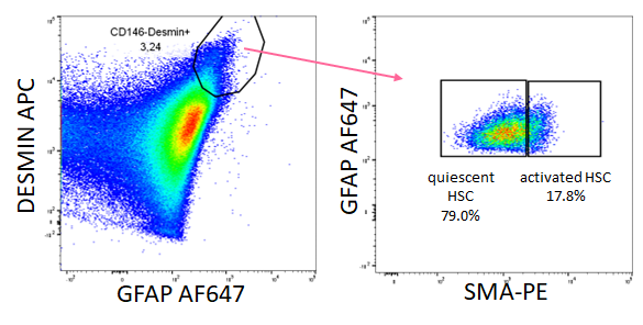
Detection by FACS of activated hepatic stellate cells (Image: María Crespo).

Methods: Male C57BL/6 J mice were subjected to either sham surgery, surgical vagotomy, or electrical vagus nerve stimulation followed by intraperitoneal injection of the TLR2 agonist zymosan. Animals were euthanized and tissues collected 12 h after injection. Samples were analyzed by qPCR, RNAseq, flow cytometry, or ELISA.

Results: Hepatic mRNA levels of pro-inflammatory mediators Ccl2, Il-1?, and Tnf-? were significantly higher in vagotomized mice compared with mice subjected to sham surgery. Differences in liver Ccl2 levels between treatment groups were largely reflected in the plasma chemokine (C-C motif) ligand 2 (CCL2) concentration. In line with this, we observed a higher number of macrophages in the livers of vagotomized mice compared with sham as measured by flow cytometry. In mice subjected to electrical vagus nerve stimulation, hepatic mRNA levels of Ccl2, Il1?, and Tnf-?, and plasma CCL2 levels, were significantly lower compared with sham. Interestingly, RNAseq revealed that a key activation marker for hepatic stellate cells (HSC), Pnpla3, was the most significantly differentially expressed gene between vagotomized and sham mice. Of note, several HSC-activation associated transcripts were higher in vagotomized mice, suggesting that signals in the vagus nerve contribute to HSC activation. In support of this, we observed significantly higher number of activated HSCs in vagotomized mice as compared with sham as measured by flow cytometry.

Conclusions: Signals in the cervical vagus nerve controlled hepatic inflammation and markers of HSC activation in zymosan-induced peritonitis.

The outcome of boosting mitochondrial activity in alcoholic liver disease (ALD) is organ-dependent

Naroa Goikoetxea-Usandizaga, Miren Bravo, Leire Egia-Mendikute, Leticia Abecia, Marina Serrano-Maciá, Rocío G Urdinguio, Marc Clos-García, Rubén Rodríguez-Agudo, Raquel Araujo-Legido, Lucía López-Bermudo, Teresa C Delgado, Sofía Lachiondo-Ortega, Irene González-Recio, Clàudia Gil-Pitarch, Ainize Peña-Cearra, Jorge Simón, Raquel Benedé-Ubieto, Silvia Ariño, Jose M Herranz, Mikel Azkargorta, Julio Salazar-Bermeo, Nuria Martí, Marta Varela-Rey, Juan M Falcón-Pérez, Óscar Lorenzo, Rubén Nogueiras, Félix Elortza Yulia A Nevzorova, Francisco J Cubero, Domingo Saura, Luis Alfonso Martínez-Cruz, Guadalupe Sabio, Asís Palazón, Pau Sancho-Bru, Natalia Elguezabal, Mario F Fraga, Matías A Ávila, Ramón Bataller, José J G Marín, Franz Martín & María Luz Martínez-Chantar.

Objective: Alcoholic liver disease (ALD) accounts for 70% of liver-related deaths in Europe, with no effective approved therapies. Although mitochondrial dysfunction is one of the earliest manifestations of alcohol-induced injury, restoring mitochondrial activity remains a problematic strategy due to oxidative stress. Here, we identify methylation-controlled J protein (MCJ) as a mediator for ALD progression and hypothesize that targeting MCJ may help recovering mitochondrial fitness without collateral oxidative damage.

Design: C57BL/6 mice (Wild-type (Wt), Mcj knockout (MCJ-KO) and Mcj liver-specific silencing (MCJ-LSS) underwent the NIAAA dietary protocol (Lieber-DeCarli diet containing 5% (vol/vol) ethanol for 10 days, plus a single binge ethanol feeding at day 11). To evaluate the impact of a restored mitochondrial activity in ALD, liver, gut, and pancreas where characterized, focusing on lipid metabolism, glucose homeostasis, intestinal permeability, and microbiota composition.

Results: MCJ, a protein acting as an endogenous negative regulator of mitochondrial respiration, is downregulated in the early stages of ALD and increases with the severity of the disease. Whole-body deficiency of MCJ is detrimental during ALD because it exacerbates the systemic effects of alcohol abuse through altered intestinal permeability, increased endotoxemia, and dysregulation of pancreatic function, which overall worsens liver injury. On the other hand, liver-specific Mcj silencing prevents main ALD hallmarks, i.e., mitochondrial dysfunction, steatosis, inflammation, and oxidative stress, as it restores the NAD+/NADH ratio and SIRT1 function, hence preventing de novo lipogenesis and improving lipid oxidation.

Conclusion: Improving mitochondrial respiration by liver-specific Mcj silencing might become a novel therapeutic approach for treating ALD.

Modulation of hypothalamic AMPK phosphorylation by olanzapine controls energy balance and body weight

Vítor Ferreira, Cintia Folgueira, Maria Guillén, Pablo Zubiaur, Marcos Navares, Assel Sarsenbayeva, Pilar López Larrubia, Jan W. Eriksson, Maria J. Pereira, Francisco Abad-Santos, Guadalupe Sabio, Patricia Rada & Ángela M. Valverde.

Second-generation antipsychotics (SGAs) are a mainstay therapy for schizophrenia. SGA-treated patients present higher risk for weight gain, dyslipidemia and hyperglycemia. Herein, we evaluated the effects of olanzapine (OLA), widely prescribed SGA, in mice focusing on changes in body weight and energy balance. We further explored OLA effects in protein tyrosine phosphatase-1B deficient (PTP1B-KO) mice, a preclinical model of leptin hypersensitivity protected against obesity.

Olanzapine injection effects on AMPK phosphorylation (Image: Cintia Folgueira).

Wild-type (WT) and PTP1B-KO mice were fed an OLA-supplemented diet (5?mg/kg/day, 7?months) or treated with OLA via intraperitoneal (i.p.) injection or by oral gavage (10?mg/kg/day, 8?weeks). Readouts of the crosstalk between hypothalamus and brown or subcutaneous white adipose tissue (BAT and iWAT, respectively) were assessed. The effects of intrahypothalamic administration of OLA with adenoviruses expressing constitutive active AMPK?1 in mice were also analyzed.

Both WT and PTP1B-KO mice receiving OLA-supplemented diet presented hyperphagia, but weight gain was enhanced only in WT mice. Unexpectedly, all mice receiving OLA via i.p. lost weight without changes in food intake, but with increased energy expenditure (EE). In these mice, reduced hypothalamic AMPK phosphorylation concurred with elevations in UCP-1 and temperature in BAT. These effects were also found by intrahypothalamic OLA injection and were abolished by constitutive activation of AMPK in the hypothalamus. Additionally, OLA i.p. treatment was associated with enhanced Tyrosine Hydroxylase (TH)-positive innervation and less sympathetic neuron-associated macrophages in iWAT. Both central and i.p. OLA injections increased UCP-1 and TH in iWAT, an effect also prevented by hypothalamic AMPK activation. By contrast, in mice fed an OLA-supplemented diet, BAT thermogenesis was only enhanced in those lacking PTP1B. Our results shed light for the first time that a threshold of OLA levels reaching the hypothalamus is required to activate the hypothalamus BAT/iWAT axis and, therefore, avoid weight gain.

Our results have unraveled an unexpected metabolic rewiring controlled by hypothalamic AMPK that avoids weight gain in male mice treated i.p. with OLA by activating BAT thermogenesis and iWAT browning and a potential benefit of PTP1B inhibition against OLA-induced weight gain upon oral treatment.

« Older posts Newer posts »

© 2026 Sabio lab

Theme by Anders Norén — Up ↑