at the CNIO

Tag: brown adipose tissue (Page 2 of 7)

Modulation of hypothalamic AMPK phosphorylation by olanzapine controls energy balance and body weight

Vítor Ferreira, Cintia Folgueira, Maria Guillén, Pablo Zubiaur, Marcos Navares, Assel Sarsenbayeva, Pilar López Larrubia, Jan W. Eriksson, Maria J. Pereira, Francisco Abad-Santos, Guadalupe Sabio, Patricia Rada & Ángela M. Valverde.

Second-generation antipsychotics (SGAs) are a mainstay therapy for schizophrenia. SGA-treated patients present higher risk for weight gain, dyslipidemia and hyperglycemia. Herein, we evaluated the effects of olanzapine (OLA), widely prescribed SGA, in mice focusing on changes in body weight and energy balance. We further explored OLA effects in protein tyrosine phosphatase-1B deficient (PTP1B-KO) mice, a preclinical model of leptin hypersensitivity protected against obesity.

Olanzapine injection effects on AMPK phosphorylation (Image: Cintia Folgueira).

Wild-type (WT) and PTP1B-KO mice were fed an OLA-supplemented diet (5 mg/kg/day, 7 months) or treated with OLA via intraperitoneal (i.p.) injection or by oral gavage (10 mg/kg/day, 8 weeks). Readouts of the crosstalk between hypothalamus and brown or subcutaneous white adipose tissue (BAT and iWAT, respectively) were assessed. The effects of intrahypothalamic administration of OLA with adenoviruses expressing constitutive active AMPKα1 in mice were also analyzed.

Both WT and PTP1B-KO mice receiving OLA-supplemented diet presented hyperphagia, but weight gain was enhanced only in WT mice. Unexpectedly, all mice receiving OLA via i.p. lost weight without changes in food intake, but with increased energy expenditure (EE). In these mice, reduced hypothalamic AMPK phosphorylation concurred with elevations in UCP-1 and temperature in BAT. These effects were also found by intrahypothalamic OLA injection and were abolished by constitutive activation of AMPK in the hypothalamus. Additionally, OLA i.p. treatment was associated with enhanced Tyrosine Hydroxylase (TH)-positive innervation and less sympathetic neuron-associated macrophages in iWAT. Both central and i.p. OLA injections increased UCP-1 and TH in iWAT, an effect also prevented by hypothalamic AMPK activation. By contrast, in mice fed an OLA-supplemented diet, BAT thermogenesis was only enhanced in those lacking PTP1B. Our results shed light for the first time that a threshold of OLA levels reaching the hypothalamus is required to activate the hypothalamus BAT/iWAT axis and, therefore, avoid weight gain.

Our results have unraveled an unexpected metabolic rewiring controlled by hypothalamic AMPK that avoids weight gain in male mice treated i.p. with OLA by activating BAT thermogenesis and iWAT browning and a potential benefit of PTP1B inhibition against OLA-induced weight gain upon oral treatment.

Targeting ERK3/MK5 complex for treatment of obesity and diabetes

Angel Loza-Valdes, Rabih El-Merahbi, Toufic Kassouf, Agnieszka Demczuk, Saskia Reuter, Jonathan Trujillo Viera, Till Karwen, Minhe Noh, Mona C. Löffler, Rafael Romero-Becerra, Jorge L. Torres, Miguel Marcos, Guadalupe Sabio, Urszula Wojda & Grzegorz Sumar.

Kinases represent one of the largest druggable families of proteins. Importantly, many kinases are aberrantly activated/de-activated in multiple organs during obesity, which contributes to the development of diabetes and associated diseases. Previous results indicate that the complex between Extracellular-regulated kinase 3 (ERK3) and Mitogen-Activated Protein Kinase (MAPK)-activated protein kinase 5 (MK5) suppresses energy dissipation and promotes fatty acids (FAs) output in adipose tissue and, therefore promotes obesity and diabetes. However, the therapeutic potential of targeting this complex at the systemic level has not been fully explored.

MK5 mRNA levels in subcutaneous adipose tissue in healthy lean subjects obese subjects.

Here we applied a translational approach to target the ERK3/MK5 complex in mice. Importantly, deletion of ERK3 in the whole body or administration of MK5-specific inhibitor protects against obesity and promotes insulin sensitivity. Finally, we show that the expression of ERK3 and MK5 correlates with the degree of obesity and that ERK3/MK5 complex regulates energy dissipation in human adipocytes.

Altogether, we demonstrate that ERK3/MK5 complex can be targeted in vivo to preserve metabolic health and combat obesity and diabetes.

Uncovering the role of p38 family members in adipose tissue physiology

Magdalena Leiva, Nuria Matesanz, Marta Pulgarín-Alfaro, Ivana Nikolić & Guadalupe Sabio.

The complex functions of adipose tissue have been a focus of research interest over the past twenty years. Adipose tissue is not only the main energy storage depot, but also one of the largest endocrine organs in the body and carries out crucial metabolic functions. Moreover, brown and beige adipose depots are major sites of energy expenditure through the activation of adaptive, non-shivering thermogenesis.

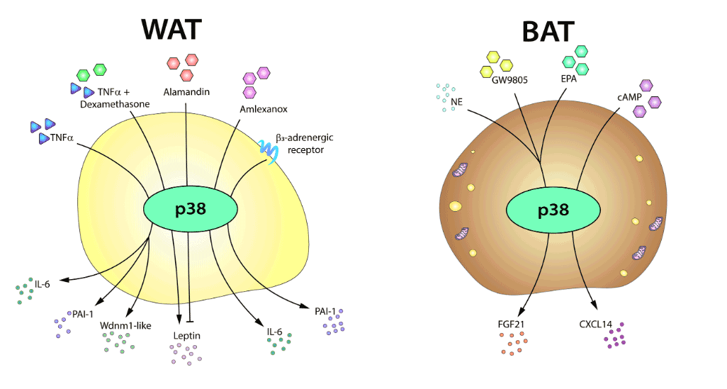
p38-mediated adipose tissue secretome
The p38-mediated adipose tissue secretome.

In recent years, numerous signaling molecules and pathways have emerged as critical regulators of adipose tissue, in both homeostasis and obesity-related disease. Among the best characterized are members of the p38 kinase family. The activity of these kinases has emerged as a key contributor to the biology of the white and brown adipose tissues, and their modulation could provide new therapeutic approaches against obesity.

Here, we give an overview of the roles of the distinct p38 family members in adipose tissue, focusing on their actions in adipogenesis, thermogenic activity, and secretory function.

« Older posts Newer posts »

© 2024 Sabio lab

Theme by Anders NorénUp ↑