Un equipo de investigadoras españolas ha descubierto dos proteínas clave para el metabolismo cardíaco después del nacimiento, un mecanismo esencial para el buen funcionamiento del corazón que, si funciona mal, puede producir daños cardíacos irreversibles, afectar a la capacidad termorreguladora y causar diabetes.
Algunos de los autores del trabajo: Alfonso Mora (izq.), Ayelén Santamans, Luis Leiva Vega, Guadalupe Sabio, José Antonio Enríquez y Bárbara González Terán.
Ayelén M. Santamans, Valle Montalvo-Romeral, Alfonso Mora, Juan Antonio Lopez, Francisco González-Romero, Daniel Jimenez-Blasco, Elena Rodríguez, Aránzazu Pintor-Chocano, Cristina Casanueva-Benítez, Rebeca Acín-Pérez, Luis Leiva-Vega, Jordi Duran, Joan J. Guinovart, Jesús Jiménez-Borreguero, José Antonio Enríquez, María Villlalba-Orero, Juan P. Bolaños, Patricia Aspichueta, Jesús Vázquez, Bárbara González-Terán, Guadalupe Sabio.
During the first weeks of postnatal heart development, cardiomyocytes undergo a major adaptive metabolic shift from glycolytic energy production to fatty acid oxidation. This metabolic change is contemporaneous to the up-regulation and activation of the p38? and p38? stress-activated protein kinases in the heart.
Cardiac fibrosis (Image: Ayelén Santamans/CNIC).
We demonstrate that p38?/? contribute to the early postnatal cardiac metabolic switch through inhibitory phosphorylation of glycogen synthase 1 (GYS1) and glycogen metabolism inactivation. Premature induction of p38?/? activation in cardiomyocytes of newborn mice results in an early GYS1 phosphorylation and inhibition of cardiac glycogen production, triggering an early metabolic shift that induces a deficit in cardiomyocyte fuel supply, leading to whole-body metabolic deregulation and maladaptive cardiac pathogenesis. Notably, the adverse effects of forced premature cardiac p38?/? activation in neonate mice are prevented by maternal diet supplementation of fatty acids during pregnancy and lactation.
These results suggest that diet interventions have a potential for treating human cardiac genetic diseases that affect heart metabolism.
Adrián Vega-Pérez, Laura H. Villarrubia, Cristina Godio, Alejandra Gutiérrez-González, Lidia Feo-Lucas, Margarita Ferriz, Natalia Martínez-Puente, Julieta Alcaín, Alfonso Mora, Guadalupe Sabio, María López-Bravo, Carlos Ardavín.
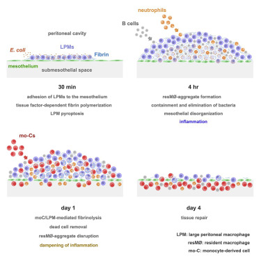
Peritoneal immune cells reside unanchored within the peritoneal fluid in homeostasis. Here, we examined the mechanisms that control bacterial infection in the peritoneum using a mouse model of abdominal sepsis following intraperitoneal Escherichia coli infection.
Whole-mount immunofluorescence and confocal microscopy of the peritoneal wall and omentum revealed that large peritoneal macrophages (LPMs) rapidly cleared bacteria and adhered to the mesothelium, forming multilayered cellular aggregates composed by sequentially recruited LPMs, B1 cells, neutrophils, and monocyte-derived cells (moCs). The formation of resident macrophage aggregates (resM?-aggregates) required LPMs and thrombin-dependent fibrin polymerization. E. coli infection triggered LPM pyroptosis and release of inflammatory mediators. Resolution of these potentially inflammatory aggregates required LPM-mediated recruitment of moCs, which were essential for fibrinolysis-mediated resM?-aggregate disaggregation and the prevention of peritoneal overt inflammation.
Thus, resM?-aggregates provide a physical scaffold that enables the efficient control of peritoneal infection, with implications for antimicrobial immunity in other body cavities, such as the pleural cavity or brain ventricles